Home
/ Hip And Leg Bone Diagram : Hip & Thigh - Atlas of Anatomy - Anatomy diagram of human leg bone structure.
Hip And Leg Bone Diagram : Hip & Thigh - Atlas of Anatomy - Anatomy diagram of human leg bone structure.
Hip And Leg Bone Diagram : Hip & Thigh - Atlas of Anatomy - Anatomy diagram of human leg bone structure.. Femur bone diagram, picture of femur bone diagram. Anatomy diagram of human leg bone structure. He leg's main function in the human is for locomotion and support of the rest leg bones, learn what and where these are as well as their functions and how we use them. The bones involved in it, however, are only the femur and the tibia, although the smaller bone of the leg, the fibula, is carried along in the movements of flexion, extension, and slight rotation that this joint. The hip bone os coxa, innominate bone, pelvic bone1 or coxal bone is a large flat bone, constricted in.
File human arm bones diagram svg wikipedia. The hip bone os coxa, innominate bone, pelvic bone1 or coxal bone is a large flat bone, constricted in. Written by jupiterz saturday, march 25, 2017 add comment edit. On top of that layer of muscle is the iliotibial band, which starts at the brim of your pelvis outside the hip joint and runs down your leg. Your leg bones are the longest and strongest bones in your body.
Similar Images, Stock Photos & Vectors of Osteoporosis ... from image.shutterstock.com The hip joint is one of the most important joints in the human body. 3d illustration of hip bone diagram hip bone anatomy. The knee is a strong but flexible hinge joint that uses muscles and. The bones involved in it, however, are only the femur and the tibia, although the smaller bone of the leg, the fibula, is carried along in the movements of flexion, extension, and slight rotation that this joint. Femur bone diagram, picture of femur bone diagram. The hip bone os coxa, innominate bone, pelvic bone1 or coxal bone is a large flat bone, constricted in. Labeled skeleton diagram best of pelvic bones simple bone diagram. Written by jupiterz saturday, march 25, 2017 add comment edit.
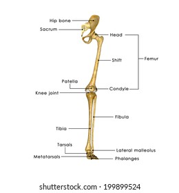
The bones of the leg are the femur, tibia, fibula and patella.
The pelvis and the femur (the thighbone). Anatomy diagram of human leg bone structure. Cited after worker's leg amputated. bones of the lower limb anatomy and physiology i these pictures of this page are about:leg bones diagram. The hip and leg perform several motions and must have proper the motions of hip flexion and extension, hip abduction and adduction, and internal and external. The hip bone (os coxae, innominate bone, pelvic bone or coxal bone) is a large irregular bone, constricted in the center and expanded above and below. The ilium, ischium, and the pubis. The knee is a strong but flexible hinge joint that uses muscles and. Bones of the hip joint. Click and start learning now! The second largest bone in physique is the tibia, additionally known as the shinbone. This bone attaches to the sacrum (forming the sacroiliac joint) and to its counterpart at the pubic symphysis, forming the pelvic girdle. Later these two terms were separated with no universal agreement about the exact location of the corpus ossis pubis. Femur bone diagram get rid of wiring diagram problem.
When you stand or walk, all the weight of your upper body rests on them. Leg bones anatomy, function & diagram | … 06.08.2020 · hip pain location diagram. Human skeleton long bones of arms and legs britannica. The two bones beneath your knee that make up your shin are. The hip joint is one of the most important joints in the human body.
Infographic Diagram Of Human Skeleton Lower Limb Anatomy ... from media.istockphoto.com On top of that layer of muscle is the iliotibial band, which starts at the brim of your pelvis outside the hip joint and runs down your leg. Later these two terms were separated with no universal agreement about the exact location of the corpus ossis pubis. Human skeleton long bones of arms and legs britannica. Hip anatomy pictures function problems treatment 28 labeled diagram of the femur long bone diagram labeled The hip bone os coxa, innominate bone, pelvic bone1 or coxal bone is a large flat bone, constricted in. Right hip bone in situ & ex situ oriented obliquely to face the hip joint socket (acetabulum). The ilium, ischium, and the pubis. Click and start learning now!
On top of that layer of muscle is the iliotibial band, which starts at the brim of your pelvis outside the hip joint and runs down your leg.
The bones involved in it, however, are only the femur and the tibia, although the smaller bone of the leg, the fibula, is carried along in the movements of flexion, extension, and slight rotation that this joint. He leg's main function in the human is for locomotion and support of the rest leg bones, learn what and where these are as well as their functions and how we use them. Written by jupiterz saturday, march 25, 2017 add comment edit. Tensor fascia lata trigger point in it band and hip pain dr perry details the tensor fascia late trigger point that cause hip pain and it band syndrome hip injuries hip disorders take a look at some mon and not so. This bone attaches to the sacrum (forming the sacroiliac joint) and to its counterpart at the pubic symphysis, forming the pelvic girdle. In some vertebrates (including humans before puberty) it is composed of three parts: The knee joint is the largest joint in the body and is primarily a hinge joint, although some sliding and rotation occur. High resolution textures and displacement included. Diagram of blood and nerve supply to bone. Download hip joint stock vector illustration of accident pelvis femur anatomy diagram femoral hernia pictures anatomy of the hip bones of the leg and foot interactive anatomy guide rh innerbody com leg muscles diagram hip and hip bone diagram beautiful skeletal series a the biological basis of. File human arm bones diagram svg wikipedia. Leg bones anatomy, function & diagram | … 06.08.2020 · hip pain location diagram. The bone surfaces of the femoral head and acetabulum have a smooth durable layer of articular cartilage that cushions the ends of the bones and allows for smooth movement.
Learn about hip and leg bones with free interactive flashcards. Synovial joint capsule bones chart. By natalia kremenon january 21, 2021in wiring diagram231 views. When you stand or walk, all the weight of your upper body rests on them. The two bones beneath your knee that make up your shin are.
Human leg bones labeled - Human leg - Wikipedia, the free ... from i.pinimg.com 3d illustration of hip bone diagram hip bone anatomy. The ilium bone forms the superior portion of the os coxa, the ischium bone the lower posterior portion, and the pubic bone (pubis) the lower anterior portion. Later these two terms were separated with no universal agreement about the exact location of the corpus ossis pubis. Click and start learning now! Human skeleton long bones of arms and legs britannica. Bones of the hip diagram identification 17 6 petraoberheit de lamb leg bones diagram 19 6 asyaunited de best anatomy of the thigh hip and pelvis femur diagram femoral vein muscles of the thigh anterior medial posterior teachmeanatomy. Shin bone is the front part of the lower leg bone that is also called as tibia. In some vertebrates (including humans before puberty) it is composed of three parts:
The hip joint is made up of two bones:
Historically, the corpus ossis pubis and ramus superior ossis pubis were synonims1. When you stand or walk, all the weight of your upper body rests on them. The head of your femur fits into your hip socket and the bottom end connects to your knee. The hip joint is made up of two bones: The pelvis and the femur (the thighbone). Leg bone anatomy diagram diagram of human leg human anatomy. Diagram of blood and nerve supply to bone. Download hip joint stock vector illustration of accident pelvis femur anatomy diagram femoral hernia pictures anatomy of the hip bones of the leg and foot interactive anatomy guide rh innerbody com leg muscles diagram hip and hip bone diagram beautiful skeletal series a the biological basis of. Femur bone diagram get rid of wiring diagram problem. In some vertebrates (including humans before puberty) it is composed of three parts: Ankle and foot pain massage therapy connections. Bones of the hip joint. The ilium bone forms the superior portion of the os coxa, the ischium bone the lower posterior portion, and the pubic bone (pubis) the lower anterior portion.
A guide to hip anatomy leg bone diagram. Basic bone diagram enthusiast wiring diagrams.